Physical Therapy Guide to Perthes Disease
Perthes disease (also known as Legg-Calvé-Perthes disease or LCPD) is a rare pediatric hip condition. It most often occurs in children 4 to 10 years old. Perthes disease begins with a disruption of blood flow to the top of the thigh bone (the femoral head), where it connects to the pelvis (hip socket). Without a blood supply, which brings oxygen and nutrients to the bone, necrosis (cell death) occurs in the vulnerable area of growing bone at the hip. Perthes disease can cause pain and limping. Perthes disease can range from mild (usually in younger children) to severe. In severe cases, the head of the femur flattens due to breakdown of bone, leading to early osteoarthritis.
The number of people with Perthes disease varies widely, with as many as 29 in 100,000 children diagnosed with the disease each year. It is more common in Caucasian children and affects boys more often than girls. The role of physical therapists in managing Perthes disease is to help children with the condition regain hip motion, decrease inflammation, limit pain, and strengthen the muscles around the hip. Physical therapists can help with activity modification to safely return children to activities they love and prevent further damage to the growing hip bone.
Physical therapists are movement experts. They improve quality of life through hands-on care, patient education, and prescribed movement. You can contact a physical therapist directly for an evaluation. To find a physical therapist in your area, visit Find a PT.
What Is Perthes Disease?
Perthes disease is a complex bone disorder that can last from several months to years. It occurs only in children and affects boys more than girls (five boys for every one girl). In 10% to15% of cases, the disease affects both hips. Children who develop the disease are often physically active and athletic. Some studies show a link between obesity in children and Perthes disease.
During childhood, bone and tissue growth are rapid. In rare cases, as children grow and for unknown reasons, the blood supply to the ball-shaped head of the femur (thigh bone) can become cut off, causing Perthes disease. This loss of blood supply affects bone growth and can cause the developing bone to lose its round shape where it connects to the hip socket.
Over time and with treatment, blood vessels regrow into the femoral head, which leads to healing and reshaping. But the healing process is slow, and the femoral head is weakened in the process. In some cases, the bone can become permanently deformed. This can result in hip disorders in adulthood, such as osteoarthritis. Early diagnosis and treatment of Perthes disease are vital to ensure long-term health.
There are four stages in Perthes disease:
Stage 1: Necrosis. Bone cells die during this initial stage. Your child may limp or avoid putting weight on the affected leg, especially after being very active. They may complain of hip, thigh, or knee pain and resist moving their hip. These symptoms usually occur without an obvious injury or incident. The initial stage may last several months.
Stage 2: Fragmentation. The body removes dead bone cells and replaces them with new, softer bone. Unfortunately, this softer bone is prone to collapse. When this happens, the head (ball) of the femur can change from a rounded shape to a more flattened shape that no longer fits in the hip socket the way it should. This can limit hip range of motion and affect walking and other activities. This stage usually spans 12 to 24 months.
Stage 3: Reossification/healing. Stronger bone begins to develop through a process called reossification. At this stage, the head of the femur begins to reform. Ideally, it returns to a normal, rounded shape, allowing unrestricted movement in the hip socket. This is usually the longest stage of healing and can take up to a few years.
Stage 4: Residual/healed. Once the bone has healed, the shape of the femoral head determines how well the joint moves and how well a child functions in everyday activities and sports. Whether the head retains close to its original round shape depends on many factors. The main factor is the child's age at the start of the disease.
Signs and Symptoms
The first signs of Perthes disease may include limping and hip or knee pain. If your child shows any of the following signs or symptoms, see a doctor or physical therapist for an evaluation:
- Limping that lasts for several days, without a known cause or injury. This is often one of the first complaints parents note. Increased activity (a long hike, running, or sports competition) may make limping worse.
- Pain on the top of the thigh, in the groin area, or in the knee. This is usually the next symptom reported. Limping may increase and your child may complain of pain when walking.
- Muscle spasms (twitches) in the thigh, hip, or buttock.
- Limited range of motion. This is especially common when rotating the foot toward the opposite leg (internal rotation) or moving the thigh away from the body (hip abduction).
- Weakness and muscle wasting (loss of muscle mass). This can occur later in the disease process. It can result in trunk sway (the trunk bends toward the affected leg) with each step.
How Is It Diagnosed?
Perthes disease is diagnosed with an X-ray or imaging of the hip and lower extremity. If your physical therapist suspects Perthes disease from their evaluation, they will refer you to a medical provider, most likely an orthopedist. A physical therapist, physician, or other health care provider will take the child's health history as part of the evaluation. During the exam, they also may:
- Observe the child walking to see if a limp is present.
- Assess pain in the hip.
- Determine whether pain is spreading to the knee or thigh.
- Measure motion at the hip to see if the child has limited movement.
- Order X-rays (which are required to confirm Perthes disease).
Typically, the child will appear otherwise well, with no general illness, trauma, or known injury. Orthopedic surgeons and radiologists rely on X-ray images to determine the stage and extent of Perthes disease. Additional tests and imaging, including MRI or bone scans, may be used to rule out other diseases or conditions.
Early diagnosis is crucial to aid full recovery. Several classification groupings exist to describe the extent of Perthes disease and predict recovery. If more than 50% of the femoral head is affected, the potential for regrowth without deformity is lower. Surgery may be needed to help preserve the femoral head's shape.
How Can a Physical Therapist Help?
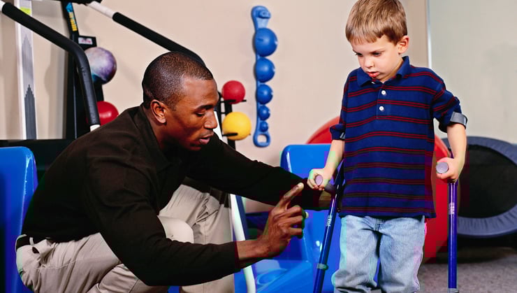
Physical therapists are important members of the health care team who work with children who have Perthes disease. Treatment is based on the severity and stage of the disease. Physical therapy can help your child maintain and improve hip range of motion, strength, and functional mobility. A physical therapist will carefully examine the child. They will develop an evidence-based exercise program designed to help restore normal hip movement and promote age-appropriate functional skills.
Your child's physical therapist will work closely with an orthopedist and pediatrician to ensure the best possible treatment outcomes.
If Surgery Is Needed
Surgery is occasionally needed to promote healing and preserve hip function. Your physical therapist will work with the surgeon's plan to help your child achieve optimal strength, mobility, and agility. A physical therapist can develop a treatment program to:
- Reduce the child's pain and inflammation.
- Help the child maintain motion in the hip.
- Educate the child and parents about safe ways to move at home and in the community that follow the surgeon's guidelines for bearing weight after the operation.
- Teach exercises for strengthening and improving cardiovascular conditioning.
- Promote safe activities during all the phases of the healing process.
Can This Injury or Condition Be Prevented?
The exact cause of Perthes disease remains unknown despite decades of study. Perthes usually occurs in only one hip but in 10%-20% of cases it affects both hips. Current research points to multiple factors that may contribute to the disease, including mechanical, genetic, and systemic conditions. Other factors linked to Perthes disease may include:
- Exposure to second-hand smoke.
- Low birth weight.
- Socioeconomic factors.
- Obesity.
- ADHD.
Noticing and identifying symptoms early is crucial to recovery. It allows treatment to begin as soon as possible and reduces or prevents problems in adulthood. Researchers continue to seek to understand the cause of Perthes disease and to improve both conservative care and surgery.
What Kind of Physical Therapist Do I Need?
All physical therapists are prepared through education and experience to treat patients with Perthes disease. However, you may want to consider:
- A physical therapist who is experienced in pediatric and orthopedic disorders. These physical therapists may work with you and your child at home, at school, or in the community, in addition to seeing them in a clinic setting.
- A physical therapist who is a board-certified clinical specialist or who has completed residency, fellowship, or training in pediatric or orthopedic physical therapy. This physical therapist has advanced knowledge, experience, and skills that may apply to rare orthopedic conditions, such as Perthes disease.
- An experienced pediatric physical therapist who also understands the importance of working with orthopedic surgeons who guide the rehabilitation plan needed to ensure the best possible outcomes for children with Perthes disease.
You can find physical therapists in your area with these credentials and clinical expertise on Find a PT, a tool built by the American Physical Therapy Association.
General tips when you're looking for a physical therapist (or any other health care provider):
- Get recommendations from family, friends, or other health care providers.
- When you contact a physical therapy clinic or home health agency for an appointment, ask about the physical therapists' experience in helping children with Perthes disease, neuromuscular disorders, or other orthopedic developmental disorders.
- Be prepared to describe your child's symptoms in as much detail as possible during your first visit. Make a note of what makes their symptoms better or worse.
The Academy of Pediatric Physical Therapy contributed to this consumer resource. It is for informational purposes only and is not intended to represent the position of APTA Pediatrics.
The American Physical Therapy Association believes consumers should have easy access to clear, reliable information that helps them make informed health care decisions and feel prepared for visits with their providers.
The following resources offer the best scientific evidence on the physical therapy treatment of Perthes disease. They cover recent research and standards of practice in the United States and globally. Whenever possible, they link to PubMed* abstracts (some of which offer free full-text access) or to other resources. You can read them to learn more or share them with your health care provider.
Braun S, Adolf S, Brenneis M, Boettner F, Meurer A. Legg-Calvé-Perthes disease-surgical treatment options. Arch Orthop Trauma Surg. 2025 Mar 12;145(1):186. Article Summary on PubMed.
American Academy of Orthopaedic Surgeons. Perthes disease. https://orthoinfo.aaos.org/en/diseases--conditions/perthes-disease. Reviewed February 2023. Accessed November 2025
Santos Santana MA, Bahiense Guimarães L, Correia Mendes L, Leal Varjao L. Effectiveness of therapeutic methods for Legg-Calvé-Perthes disease according to staging, limits of conservative treatment: a systematic review with meta-analysis. Orthopedic Reviews. 2024;16. Article Summary on PubMed.
US Library of Medicine, Medline Plus. Legg-Calve-Perthes disease. https://medlineplus.gov/genetics/condition/legg-calve-perthes-disease/. Accessed July 30, 2021.
National Organization for Rare Disorders. Legg-Calvé-Perthes disease: symptoms of Legg-Calvé-Perthes disease. Updated 2016. Accessed June 2, 2021.
Mazloumi SM, Ebrahimzadeh MH, Kachooei AR. Evolution in diagnosis and treatment of Legg-Calve-Perthes disease. Arch Bone Jt Surg. 2014;2(2):86-92. Article Summary on PubMed.
*PubMed is a free public website run by the National Library of Medicine. It allows people to access summaries and references from health research articles published in scientific journals, including those indexed in the MEDLINE database.
Expert Review:
Nov 16, 2025
Revised:
May 14, 2026
Content Type: Guide
Perthes Disease
PT, DPT, board-certified clinical specialist in pediatric physical therapy
PT, ScD, board-certified clinical specialist in pediatric physical therapy
Ann Smith
PT, DPT, board-certified clinical specialist in orthopedic and pediatric physical therapy